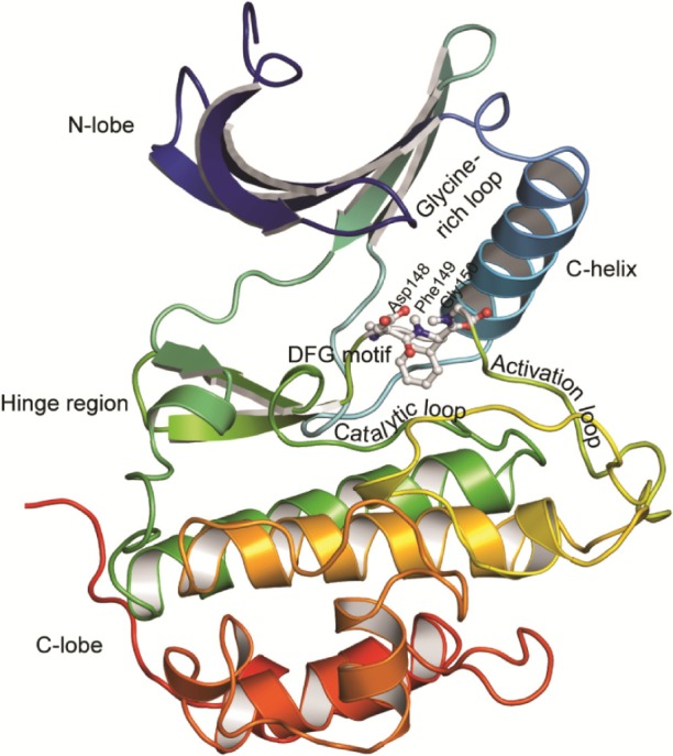
Figure 2.

Structure of serine/threonine kinases.
Notes: The protein is shown in cartoon representation and colored in rainbow colors with violet at the N-terminus and red at the C-terminus of the structure. The N- and C-lobes with the connecting hinge region are indicated. The catalytic loop, activation loop, glycine-rich loop, C-helix, and the DFG motif are labeled. The Chk1 protein structure (PDB ID: 1ZYS) belonging to the CaMK family of serine/threonine protein kinases was used to generate this image.
Abbreviations: C-lobe, C-terminal lobe; DFG, Asp–Phe–Gly; N-lobe, N-terminal lobe; PDB, Protein Data Bank.
