Figure 3.
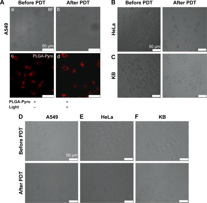
Cell morphological changes observed by microscopy during Pyro-loaded PEG-PLGA NP-based and free Pyro-based PDT.
Notes: Microscopy images of (A) A549 cells. BF image of A549 cells before Pyro-loaded NP-based PDT (a). BF image of A549 cells after Pyro-loaded NP-based PDT (b). Fluorescence images of A549 cells before Pyro-loaded NP-based PDT (c). Fluorescence images of A549 cells after Pyro-loaded NP-based PDT (d). (B) Pyro-loaded NP-based PDT on HeLa cells and (C) Pyro-loaded NP-based PDT on KB cells. (D) Free Pyro-based PDT on A549 cells. (E) Free Pyro-based PDT on HeLa cells. (F) Free Pyro-based PDT on KB cells.
Abbreviations: Pyro, pyropheophorbide-a; PEG, polyethylene glycol; PLGA, poly(lactic-co-glycolic acid); NP, nanoparticle; PDT, photodynamic therapy; BF, bright field.

