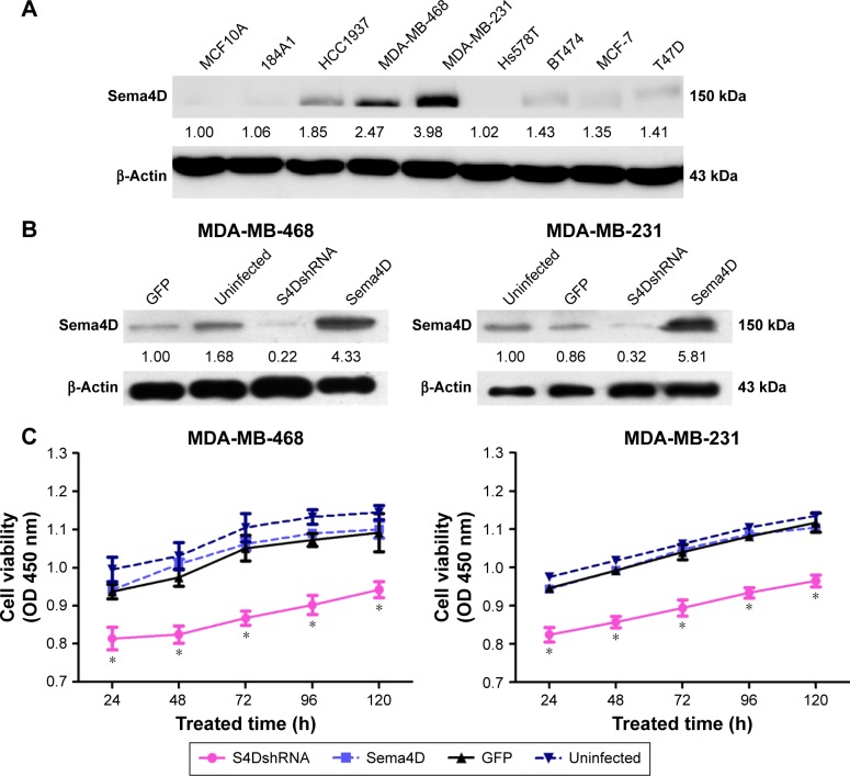
Figure 1.
The expression of Sema4D in seven breast cancer cell lines and in normal human breast epithelial cell lines.
Notes: (A) Sema4D protein levels were detected by Western blotting; β-actin was used as a loading control (lower panels). The following cell lines were analyzed: HCC1937, MDA-MB-231, MDA-MB-468, Hs578T, BT474, MCF-7, T47D, and the normal human breast epithelial cell lines MCF10A and 184A1. Protein levels are quantified below each blot as the fold increase relative to MCF10A cells. Bars indicate the means of three individual experiments ± standard errors. (B) Western blotting analysis of Sema4D in the indicated cell lines; β-actin was used as the loading control. Protein levels are quantified below each blot as the fold increase relative to loading controls. Each cell line was separately infected by control lentivirus-expressing enhanced GFP, lentivirus coding for Sema4D shRNA, and lentivirus coding for Sema4D. The following cell lines were included in the analysis: MDA-MB-231 and MDA-MB-468 (*P<0.05, relative to both the uninfected and GFP groups). (C) The proliferation of Sema4D in MDA-MB-231 and MDA-MB-468 cells was detected using the MTT assay.
Abbreviations: Sema4D, semaphorin 4D; shRNA, short hairpin RNA.

