Figure 2.

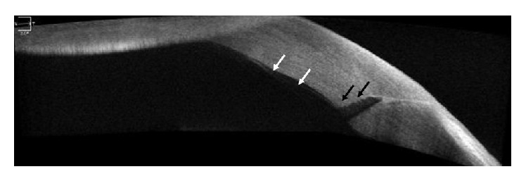
A TD AS-OCT image demonstrating localized Descemet's membrane detachment (white arrow) adjacent to the wound and internal wound gaping (black arrow) that was not detected in slit-lamp examination.

A TD AS-OCT image demonstrating localized Descemet's membrane detachment (white arrow) adjacent to the wound and internal wound gaping (black arrow) that was not detected in slit-lamp examination.