
Figure 1.
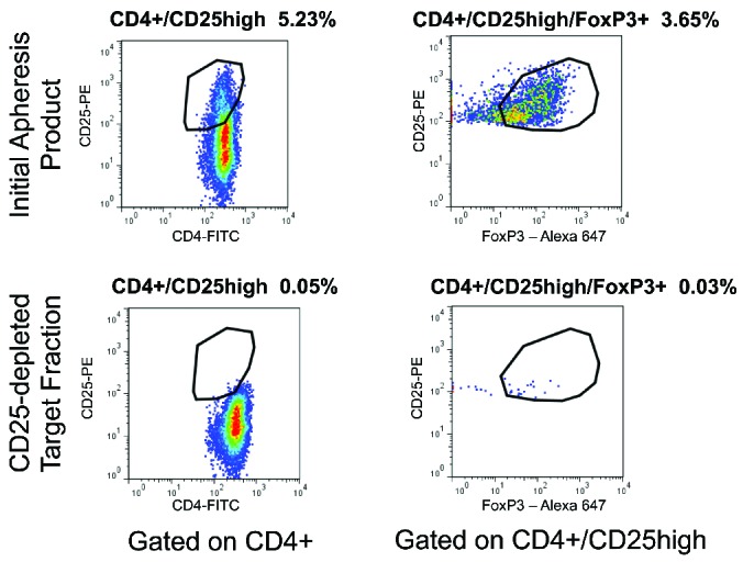
Flow cytometric analysis of CD4+CD25+FoxP3+ cells confirms Treg depletion. The initial apheresis product (top plots) and target fraction after CD25 depletion using the CliniMACS system (bottom plots) were subjected to extracellular staining for CD4 and CD25 expression and intracellular staining for FoxP3 protein. As shown for a representative DLI product, gating on CD45+CD4+ cells demonstrated reduction in cells expressing high levels of CD25 (left plots). Gating on CD4+CD25high cells demonstrated reduction in cells expressing FoxP3 (right plots) after the depletion process.