
Figure 3.
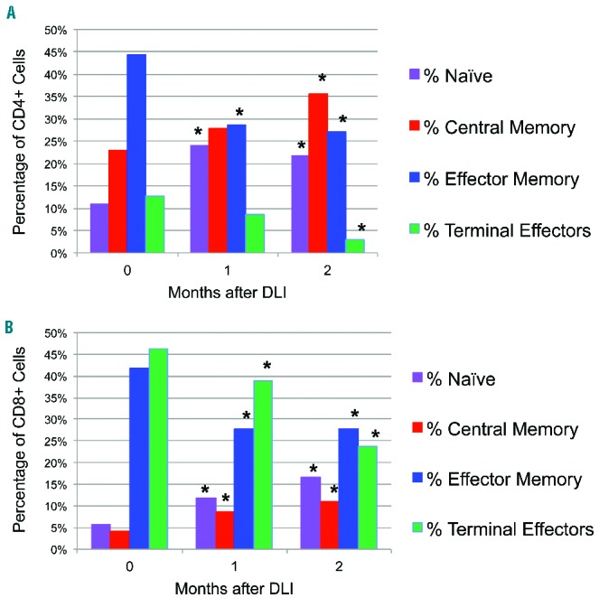
Naïve and central memory cells increase within CD4+ and CD8+ T-cell compartments after CD25/Treg-depleted DLI. Peripheral blood mononuclear cells drawn at indicated times were stained for extracellular markers and analyzed by flow cytometry. (A) Changes in median distribution of CD4+ T cells among all subjects: the percentage of CD3+CD4+ T cells in each subset just prior to (0 months) or in the first 2 months after DLI is indicated. Panels reflect percentage of naïve cells (CD45RO-CD62L+), central memory cells (CD45RO+CD62L+), effector memory cells (CD45RO+CD62L−), and terminal effectors (CD45RO−CD62L−). *Indicates P<0.05 for the comparison between baseline (month 0) and the indicated time point. (B) Changes in median distribution of CD8+ T cells. Individual CD8+ subsets and significant differences among all subjects after CD25/Treg-depleted DLI are displayed as detailed above.