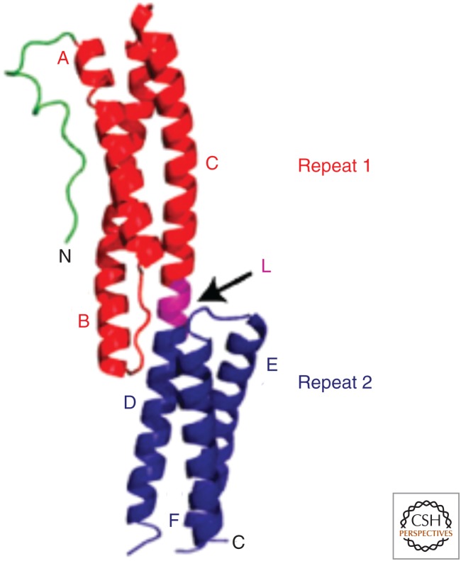
Figure 2.
Ribbon diagram illustrating the structure of two spectrin repeats (SRs) in the plakin domain of bullous pemphigoid antigen 1 (BPAG1/dystonin), which is a member of the spectrin superfamily. Depicted is a loop-like region (green) at the far amino-terminal end, followed by a pair of SRs 1 and 2 (red and blue, respectively) arranged in tandem and connected by a linker region (L, purple) that is also helical in nature. Also labeled are the component helices A–C and D–F. (Reprinted from Jefferson et al. 2004.)

