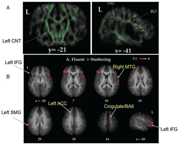
Figure 2.
(A) Differences in white matter integrity as assessed through tract-based spatial statistics (TBSS) of diffusion tensor imaging data. TBSS allows whole-brain comparisons of measures of white matter integrity. Here, 8- to 12-year-old boys who stutter exhibited significantly decreased white matter integrity in the superior longitudinal fasciculus, underlying the rolandic operculum (RO), and the bilateral corticonuclear tracts (CNT). (B) Differences in gray matter volume between boys who do and boys who do not stutter. The same group of 8- to 12-year-old participants were examined for differences in gray matter volume (GMV) across the whole brain. Red blobs show areas where boys who stutter exhibited less GMV compared with age-matched peers. These regions included the left inferior frontal gyrus (IFG), right superior and middle temporal gyrus (MTG), left supramarginal gyrus (SMG), and anterior cingulate cortex (ACC)/supplementary motor area.29

