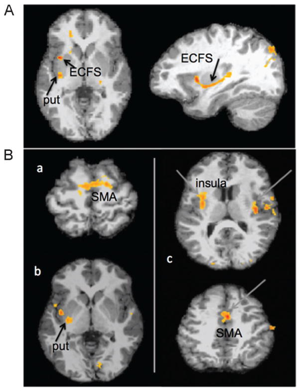
Figure 3.
(A) Areas where the probability of white matter tracts were decreased in children who stutter, when starting point of the white matter tracking was in the left posterior superior temporal gyrus (pSTG). Children who stutter showed decreased probability of white matter tracts in the putamen (put) and inferior frontal gyrus, via the external capsule fiber system (ECFS). (B) Differences in brain activity patterns in children who stutter when they were at rest. Resting state functional magnetic resonance imaging data were analyzed to compare correlated brain activity patterns (which indicate areas that “talk with each other,” or the presence of functional connectivity between those regions) between the stuttering and control groups. (a) When correlated brain activity with the left putamen was examined, the supplementary motor area (SMA) had significantly greater correlation with putamen in the typically speaking children compared with children who stutter. (b) When correlated activity with the left SMA was examined, the putamen insula showed significantly heightened correlated activity with the SMA in the typically speaking controls compared with children who stutter. (c) When correlated activity with the left pSTG was examined, the bilateral insula, STG, and SMA regions showed greater correlation in typically speaking controls compared with children who stutter.43

