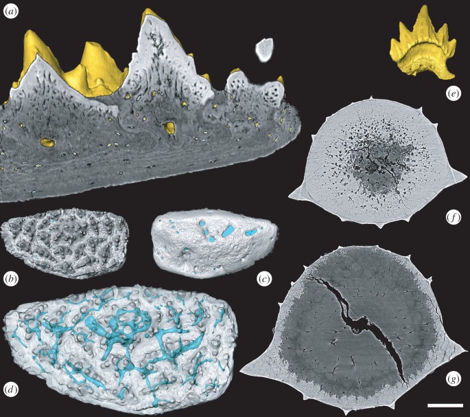
Figure 1.
Surface rendering and virtual thin-section of Romundina supragnathal (NRM-PZ P.15956, a–d) and Scyliorhinus canicula (BRSUG 29402) teeth (e–g). Romundina pulp cavities in transverse section (a) and vascular system in oral (b), dorsal (c) and oral transparent view (d). Scyliorhinus tooth (e) enameloid layer with radial structures (f,g). Scale bar represents 50 µm in (a), 417 µm in (b,c), 240 µm (d), 480 µm (e), 37 µm (f) and 45 µm (g).

