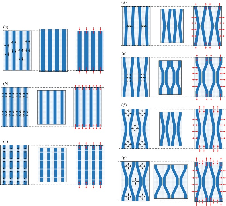
Figure 3.
Abstract representation of each model: (a) lateral crystal growth, (b) amorphous cellulose at the surface of microfibrils, (c) amorphous cellulose in series with the microfibrils, (d) active binding of microfibrils, (e) entrapment of matrix material during cellulose aggregation, (f) drying of the G-layer during maturation, (g) matrix swelling in a connected cellulose network. For each model, cell wall material is represented in three states: (left) state before maturation, (middle) virtual state of deformation if the volume was free to strain, (right) in situ state of stress. Black arrows inside constituents represent their initial tendency to shrink or swell (convergent arrows indicate shrinkage and divergent arrows indicate swelling). Red arrows represent the final state of stress at the border of the elementary volume (convergent arrows indicate compression and divergent arrows indicate tension). For a complete description of the mechanisms, see text.

