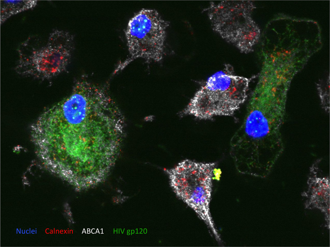
Figure 3. A 2D image of monocyte-derived macrophages infected with HIV-1.
Peripheral blood monocytes were differentiated into the macrophages and infected with VSV-G pseudotyped HIV-1 NL4-3. HIV-1 envelope protein was stained using anti-HIV gp120 sheep serum followed by Alexa Fluor® 488 Donkey Anti-Sheep IgG (H+L) Antibody (green). Cellular proteins Calnexin and ABCA1 were visualized using mouse monoclonal anti-Calnexin-ER membrane marker antibody followed by goat anti-mouse DyLight 550 antibody (red) and by anti-ABCA1 rabbit polyclonal antibody followed by goat anti rabbit Alexa-Fluor 647 antibody (white), respectively. Cellular nuclei were stained by DAPI dilactate (blue). Abundance of ABCA1 is reduced in HIV-infected macrophages.

