
Figure 3. Correlation of WSS with tissue histopathology.
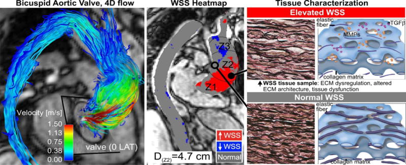
Eccentric transvalvular BAV flow (left) exposes aortic wall regions exposed to elevated WSS (middle, red region) which exhibit abnormal tissue metrics of aortopathy (right)(71).
Official websites use .gov
A
.gov website belongs to an official
government organization in the United States.
Secure .gov websites use HTTPS
A lock (
) or https:// means you've safely
connected to the .gov website. Share sensitive
information only on official, secure websites.
Eccentric transvalvular BAV flow (left) exposes aortic wall regions exposed to elevated WSS (middle, red region) which exhibit abnormal tissue metrics of aortopathy (right)(71).