Figure 6.
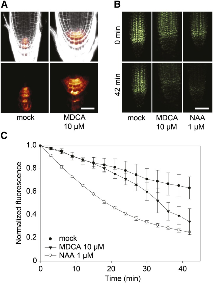
Activity of auxin reporters in MDCA-treated Arabidopsis seedlings. A, Confocal images of MDCA-induced activation of the auxin-responsive DR5 promoter in the primary root tip of pDR5rev:GFP seedlings. Seedlings were germinated 7 d on 0.5 × MS-medium before being transferred to 0.5 × MS-medium supplemented with 10 μM MDCA for 5 d (n = 5; scale bar: 35 μm). PI was used as counterstain to visualize the cell walls. Color-code depicts (red; low to white; high) pDR5rev:GFP signal intensity. B, Confocal images of DII-VENUS degradation in the primary root tip of DII-VENUS-YFP seedlings. Seedlings were germinated 7 d on 0.5 × MS-medium before being transferred to 0.5 × MS-medium with 10 μM MDCA or 1 μM NAA. Individual root tips were imaged at the onset of the experiment and after 42 min (n = 3; scale bar: 50 μm). C, Time-course of DII-VENUS fluorescence in the primary root tips of seedlings grown as described above. Fluorescence was quantified every 3 min over a 42-min interval. The fluorescence was normalized against the initial value for each treatment. Error bars represent sds.

