Figure 7.
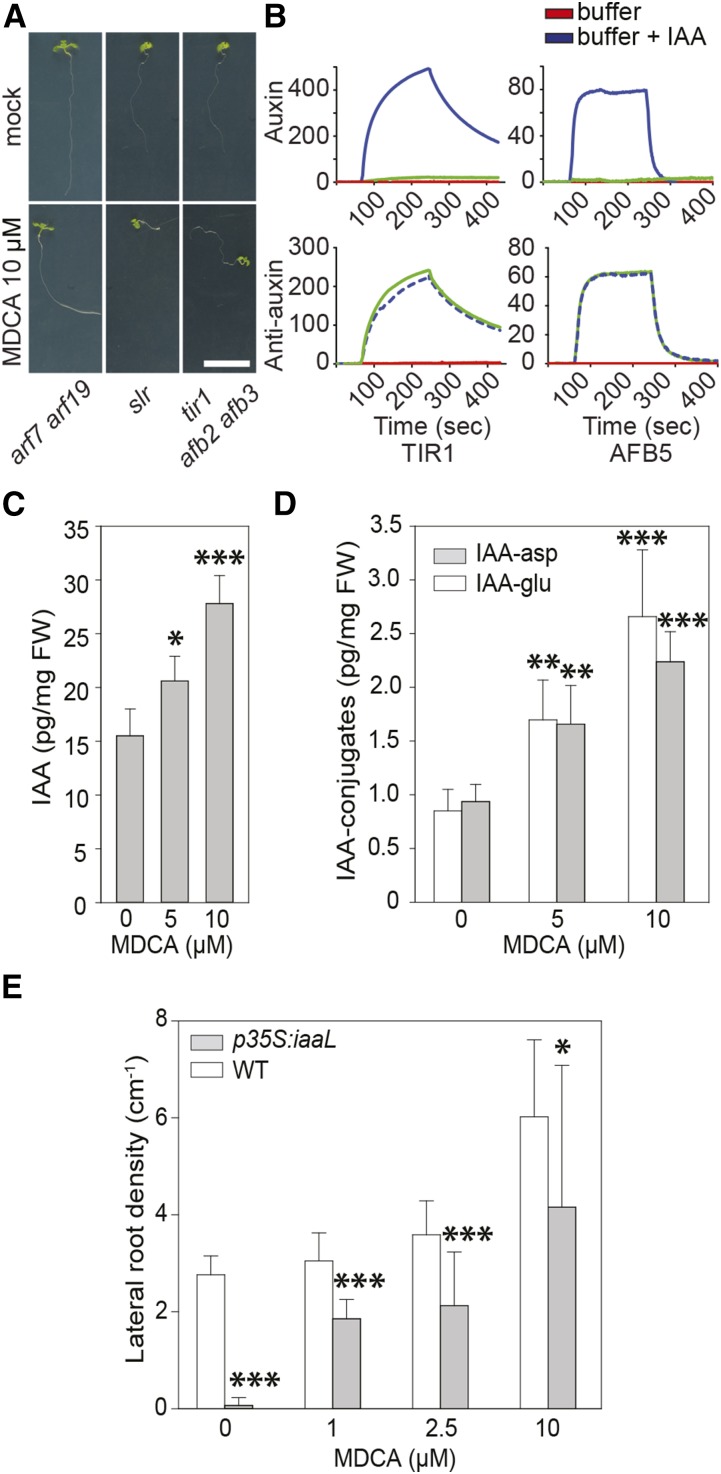
The importance of auxin in MDCA-mediated developmental defects in seedlings. A, Root phenotype of arf7 arf19, slr, and tir1 afb2 afb3 mutant seedlings (12 DAG) grown on 0.5 × MS-medium supplemented with 10 μM MDCA (n > 25; scale bar: 1 cm). B, SPR sensorgrams showing the auxin-dependent interaction between TIR1 or AFB5 with IAA7/14 DII. Each sensorgram shows the binding with IAA (blue), an auxin-free injection (red), plus the data for MDCA (green). For auxin activity assays, MDCA (50 μM) was mixed with TIR1 or AFB5 prior to injection over DII peptide. For antiauxin assays, MDCA (50 μM) was mixed with TIR1 or AFB5 plus 5 μM IAA prior to injection. Dashed lines were used to increase the visibility. (C and D) Free IAA levels and IAA-amino acid conjugate (IAA-Asp and IAA-Glu) levels of seedlings (12 DAG) grown on 0.5 × MS-medium supplemented with different concentrations of MDCA. E, Each biological replicate represents 10 seedlings that were pooled and analyzed (n = 6). Error bars represent sds and asterisks were used to indicate statistically significant differences between wild type and p35S:iaaL at a given concentration as determined by Dunnett’s test P values: *P < 0.05, **P < 0.001, ***P < 0.0001.

