Figure 1.
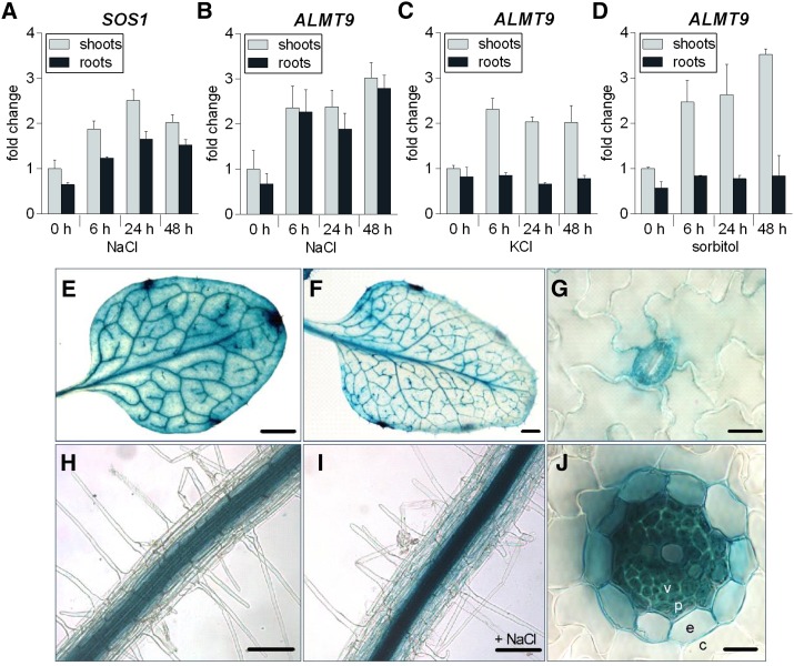
Transcriptional regulation and expression pattern analysis of ALMT9. A to D, qRT-PCR analysis of SOS1 (A) and ALMT9 (B–D) expression in hydroponically grown wild-type shoots and roots after the application of 100 mm NaCl (A and B), 100 mm KCl (C), and 200 mm sorbitol (D) for 0, 6, 24, and 48 h. The data were normalized to expression levels in shoots prior to treatment (0 h). ACT2 served as a reference gene. Data are means ± sd of n = 3 biological replicates. E to J, ALMT9 expression pattern revealed by histochemical localization of GUS activity directed by the ALMT9 promoter. E, Mesophyll cell and vasculature expression in the third rosette leaf. F, Vasculature expression in the sixth rosette leaf. G, Expression in guard cells. H, Expression in root stelar cells. I, Expression in response to 100 mm NaCl for 24 h was enhanced but remained restricted to the root stele. J, In cross-sections of roots, no expression was detected in cortex cells (c), but in the endodermis (e), the pericycle (p), and the vasculature (v). Scale bars represent 0.2 mm in E and F, 10 µm in G and J, and 100 µm in H and I.

