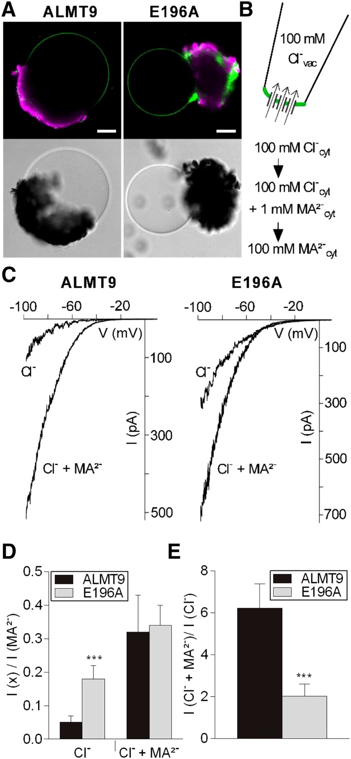
Figure 3.
Electrophysiological properties of the mutant channel ALMT9E196A. A, Fluorescence and transmission images of vacuoles released from lysed tobacco protoplasts that transiently overexpress ALMT9-GFP (left) and ALMT9E196A-GFP (right). Auto-fluorescence of chloroplasts is shown in magenta. Scale bars = 20 µm. B, Patch-clamp experimental procedure. Vacuoles were patched in excised cytosolic-side-out configuration under symmetric ionic conditions (100 mm Cl−vac/ 100 mm Cl−cyt). The cytosolic buffer was sequentially exchanged (100 mm Cl−; 100 mm Cl− + 1 mm MA2−; 100 mm MA2−) on the same membrane patch. C, Representative currents of ALMT9 (left) and ALMT9E196A (right) in presence of 100 mm Cl− (Cl−) and 100 mm Cl− + 1 mm MA2− (Cl− + MA2−) in the cytosolic buffers. Currents were evoked by a 2.5-s voltage ramp ranging from +40 mV to −100 mV. D, Relative Cl− and Cl− + MA2− currents mediated by ALMT9 and ALMT9E196A. Currents were normalized to the current amplitude measured at −100 mV in presence of 100 mm MA2− in the cytosolic solution. E, Level of MA2−-activation (ICl− + MA2−/ ICl−) of ALMT9- and ALMT9E196A-mediated Cl− currents at −100 mV. Data are means ± sd. Asterisks indicate statistically significant differences between ALMT9 (n = 5) and ALMT9E196A (n = 6) currents (*P < 0.05, **P < 0.01, ***P < 0.001; two-tailed Student’s t test).

