Figure 5.
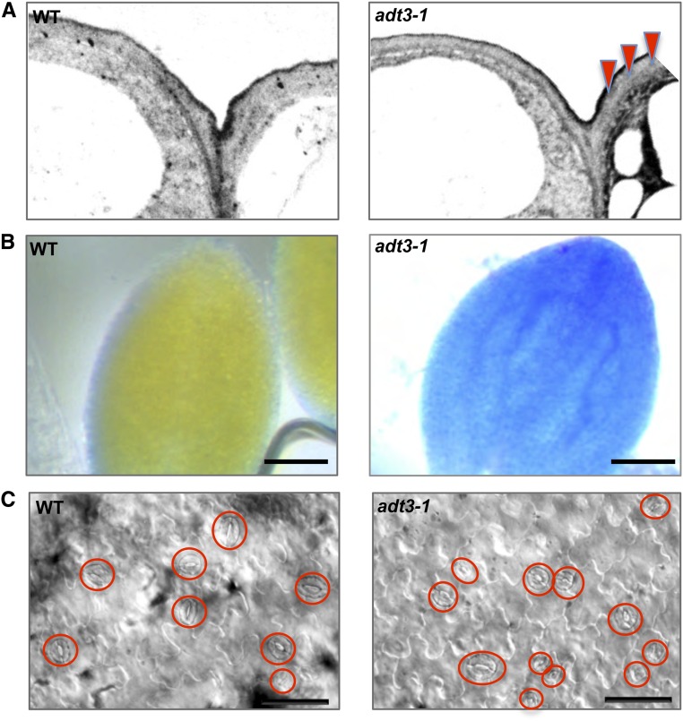
Defective cuticle formation, altered permeability, and aberrant patterning of guard cells in adt3-1 seedlings. A, Ultrastructure of the cuticle of adt3-1 epidermis. TEM micrographs of wild-type (WT; top) and adt3-1 (bottom) epidermis show the deposition of electron-opaque material within the adt3-1 cell wall (red triangles). B, Increased permeability of adt3-1 epidermis under hyposmotic treatment. TB penetration is shown in dark-grown, 6-d-old wild-type (top) and adt3-1 (bottom) seedlings after 48 h of incubation in sterile water. Bars = 50 μm. C, Light-grown (16 h light:8 h dark), 6-d-old adt3-1 seedlings exhibit abnormal stomatal development; guard cells in the focal plane are circled. Bars = 25 μm.

