Figure 6.
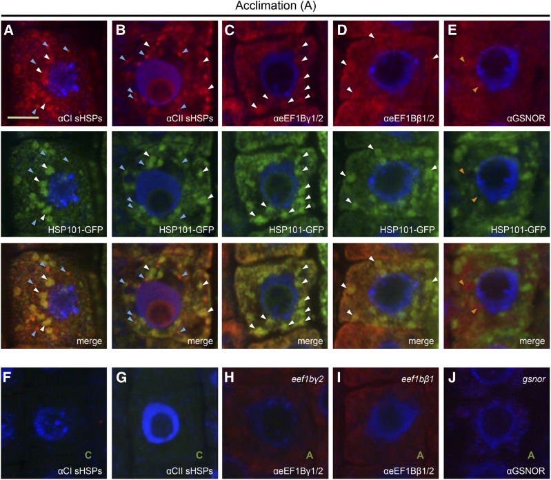
CI and CII sHSPs and eEF1B β- and γ-subunits colocalize with HSP101-GFP. Arabidopsis roots expressing HSP101-GFP were heat acclimated (90 min, 38°C plus 2 h, 21°C), fixed, and processed for immunolocalization using the indicated polyclonal antibodies. HSP101-GFP was localized based on GFP fluorescence, and all other proteins were detected using secondary antibodies coupled to Alexa 594. A, CI sHSPs localize to small bright cytosolic foci (blue arrowheads) and to larger aggregates (white arrowheads) that colocalize with HSP101-GFP. B, CII sHSP localization was similar to CI sHSPs, but there are a larger number of small bright cytosolic foci with CII sHSPs (blue arrowheads). C and D, eEF1Bγ and eEF1Bβ show partial colocalization with HSP101-GFP-containing aggregates. E, GSNOR is more uniformly distributed in the cell and did not colocalize with HSP101-GFP-containing aggregates (orange arrowheads). F and G, Plants kept in control conditions were treated similarly to those in A and B and did not show any detectable signal in the red and green channels, verifying the specificity of the signal. H and I, The eEF1Bγ2 and eEF1Bβ1 T-DNA insertion lines displayed a reduced signal with the corresponding antibodies. J, The gsnor mutant did not show any detectable signal when using the GSNOR antibody, verifying the specificity of the signal.

