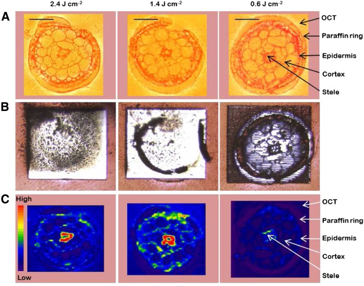
Figure 3.
Optimization of LA-ICP-MS settings in order to obtain a quantitative ablation and to achieve the best possible spatial resolution when analyzing cross sections from Arabidopsis roots. All three cross sections were 16 µm thick and were analyzed with decreasing energy levels (left to right) and otherwise identical settings. The energy levels in the laser beam were 2.36, 1.4, and 0.59 J cm−2, respectively. A shows bright-field microscopy images, B shows microscopy images taken after the ablation, and C shows the distribution and ion intensity of K, analyzed as 39K, with LA-ICP-MS. The red-to-blue color spectrum in C represents high to low intensities, respectively (range, 2,000–80,000 counts). Bars = 50 µm.

