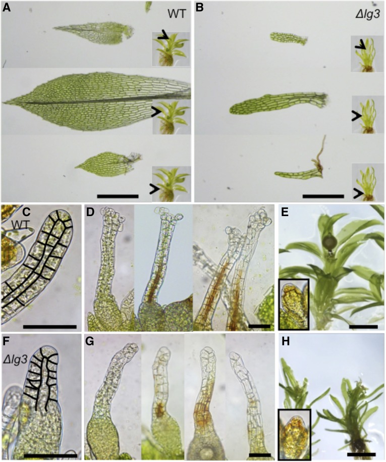
Figure 5.
Phyllid and reproductive organ morphology in the wild type (WT) and dek1Δlg3. A, Wild-type phyllids isolated from the basal, middle, and apical parts of the gametophore. The positions of the isolated phyllids on gametophores are marked by arrowheads in the insets. B, dek1Δlg3 phyllids isolated from the basal, middle, and apical parts of the gametophore. The positions of the isolated phyllids on gametophores are marked by arrowheads in the insets. C, Young wild-type archegonium with highlighted cell walls in the neck. D, Examples of wild-type archegonia at the later stages of development. Note the open apices and brown pigmentation marking the neck canal. E, Wild-type gametophore with sporophyte. Antheridium is shown in the inset. F, Young dek1Δlg3 archegonium with highlighted cell walls in the neck. G, dek1Δlg3 archegonia at the later stages of development. Note the closed and deformed necks and patchy distribution of the brown pigmentation that reflects defects in the neck canal differentiation. H, dek1Δlg3 gametophores cultivated under the sporogenesis conditions. Sporophytes are not formed. Antheridium is shown in the inset. Bars = 500 μm (A and B), 50 μm (C, D, F, and G), and 1 mm (E and H).

