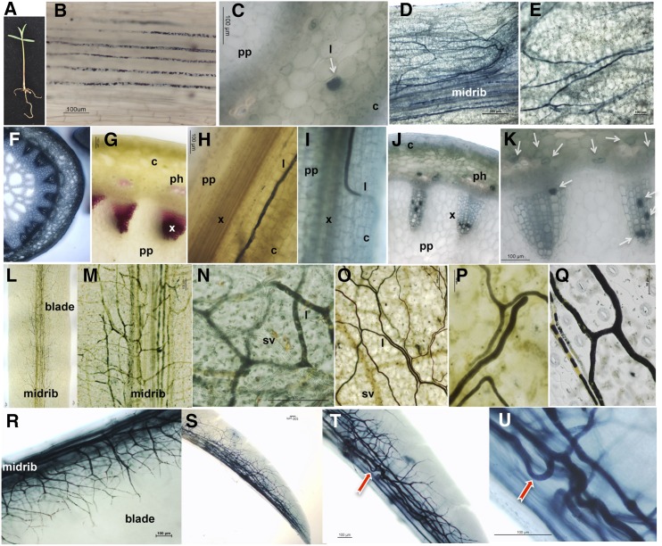
Figure 1.
Distribution pattern of laticifer cells in E. lathyris intact plant structures as revealed by whole-mount staining with Sudan Black B. A, Seedling, showing the first pair of true leaves, employed to identify laticifer cells as in B to E. B, A sector of the hypocotyl showing rows of laticifers running in parallel along the hypocotyl. C, Cross section of a hypocotyl region with laticifer cells (marked by a white arrow) specifically stained for isoprenoids. D and E, Longitudinal sector of a whole-mount stained cotyledon, along the lamina, in a region proximal to the node (D), where longitudinal laticifers cells concentrate along the midrib and in a distal region from the node and midrib (E), where laticifers appear scattered. F and G, Cross section of the stem stained with toluidine blue (F) and phloroglucinol (G), where the lignified xylem poles become specifically stained in red. H and I, Close-up of a whole-mount preparation of the stem showing an ascending laticifer running parallel to one of the poles of the vascular cylinder (H) or curving toward the vasculature from the cortex in search of a xylem pole (I). J and K, Close-up of cross sections of the stem showing some laticifer cells occupying the internal cavity of the hollow cylinder of different tracheary elements. L, Sector of a blade from a whole-mount stained fully expanded leaf. M, Magnification of a sector showing the midrib of the leaf blade and the abundant presence of laticifer cells. N and O, Magnification of leaf blade sectors showing the different dispositions and distribution patterns of laticifer cells. P and Q, Magnification of leaf blade laticifer cells showing characteristic Y- and H-bifurcation patterns. R to U, Whole-mount preparation of an emerging leaf close to the apical meristem, with predominant distribution of laticifer cells close to the midrib (R) and details of a sector close to the leaf tip at different magnifications (S–U). c, Cortex; l, laticifer; ph, phloem; pp, pith parenchyma; sv, secondary vasculature; x, xylem.

