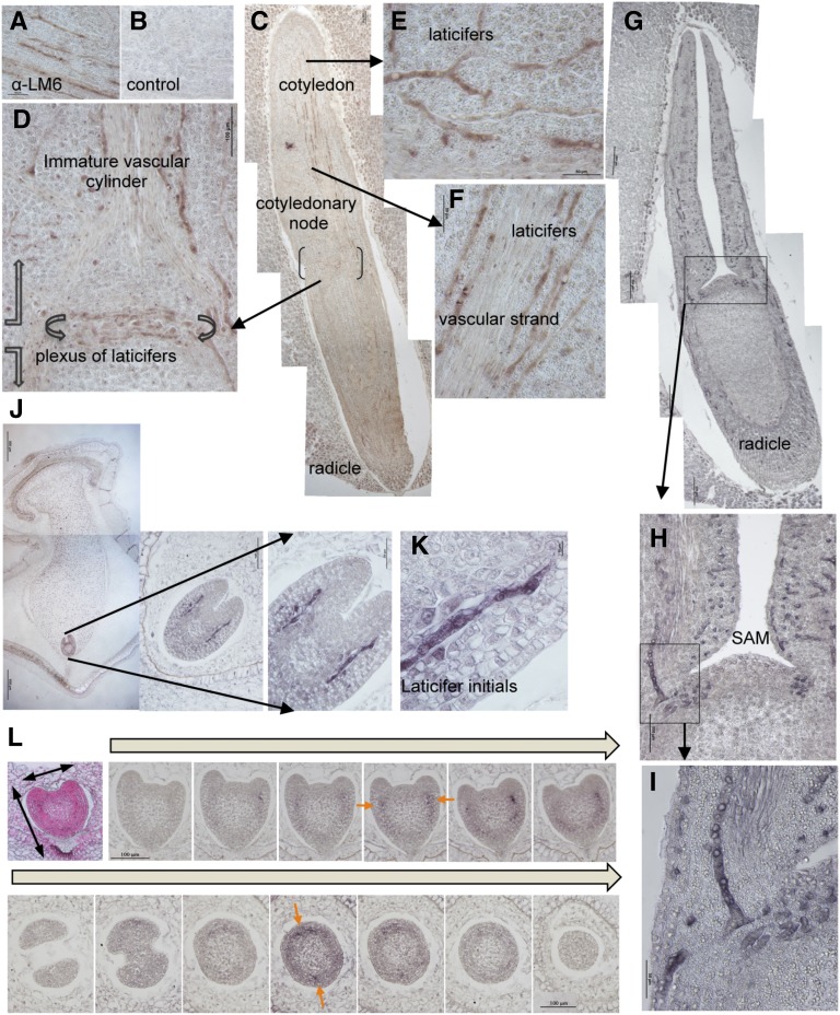
Figure 3.
Organization and ontogeny of the laticiferous system in the E. lathyris embryo. A and B, Comparative immunohistochemical staining of E. lathyris embryo sections with LM6 (A) and a nonspecific (B) antibody revealed the specificity of LM6 to immunodecorate laticifer cells in the embryo. C, Sagittal section along the longitudinal axis of the mature embryo revealed with LM6 allowed identifying laticifer cellular structures along the embryo axis. D, Magnification of the sector shown in C showing the ring of interwoven laticifer cells (plexus) in the cotyledonary node and the ascending row of laticifers parallel to the immature vascular strand in the cotyledon. E and F, Details of branches of laticifer cell structures extending laterally (E) in the cotyledon tissues or extending upwards and parallel to the immature vascular strand (F). G–I, Frontal view of a mature embryo section immunodecorated with LM6 (G; magnification details (H and I) allowed identification of laticifer branches ascending into the cotyledons. Observed that the sam appears devoid of laticifer strands. J, Detection of elongated laticifer initials in immature embryos (i.e. heart stage) as embedded in the seed. The laticifers appeared immunodecorated at the base of the emerging cotyledons. K, Detail of a laticifer initial terminating as a narrow tip with acuminate ends. L, Detection of earliest laticifer appearance detected using LM6 antibodies at late globular stages of embryo development. Serial longitudinal sectioning (top) and cross-sectioning (bottom) of embryos revealed the presence of a single pair of nonelongated initials, marked with orange arrows and discernible at the time primordia of cotyledons start to form in the immature embryo.

