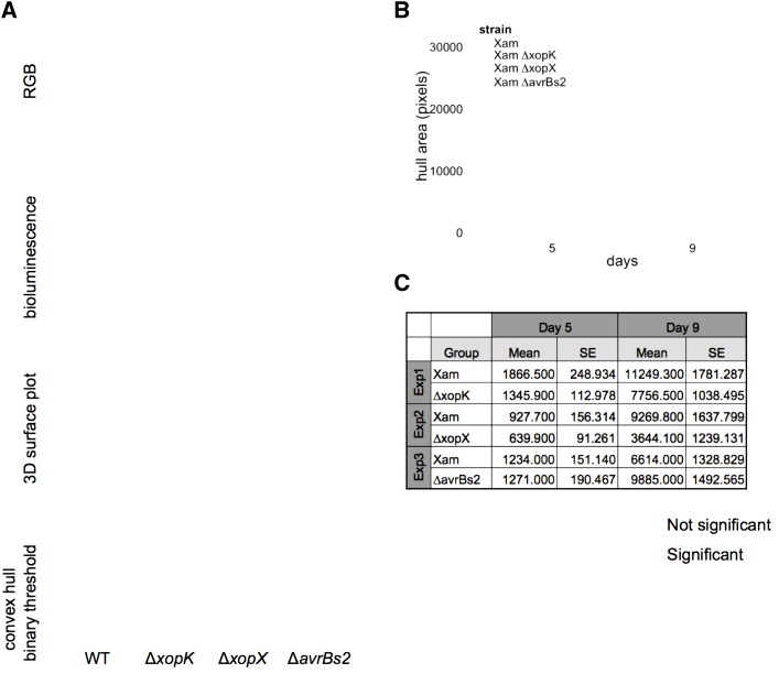
Figure 3.
Bioluminescence imaging of Xam spread in cassava leaves. A, The bioluminescence reporter pLUX plasmid was introduced into Xam strains. Leaves were inoculated with bacterial solutions (OD600 = 0.01) using syringe infiltration. RGB images of inoculated leaves reveal symptom development. Bioluminescence was visualized in a dark chamber with a 5–10 min exposure. Image processing was performed with ImageJ to select the area of bioluminescence and the convex hull of the resulting shapes were analyzed. B, Representative quantification of convex hull for wild-type Xam and three mutants. Additional replicate experiments are shown in Supplemental Figure S4. C, Results of generalized linear mixed model analysis of convex hull area, combining data from all replicate experiments. Combined estimated means and se are presented, as well as the difference between the means and the P values for each pairwise statistical contrast.

