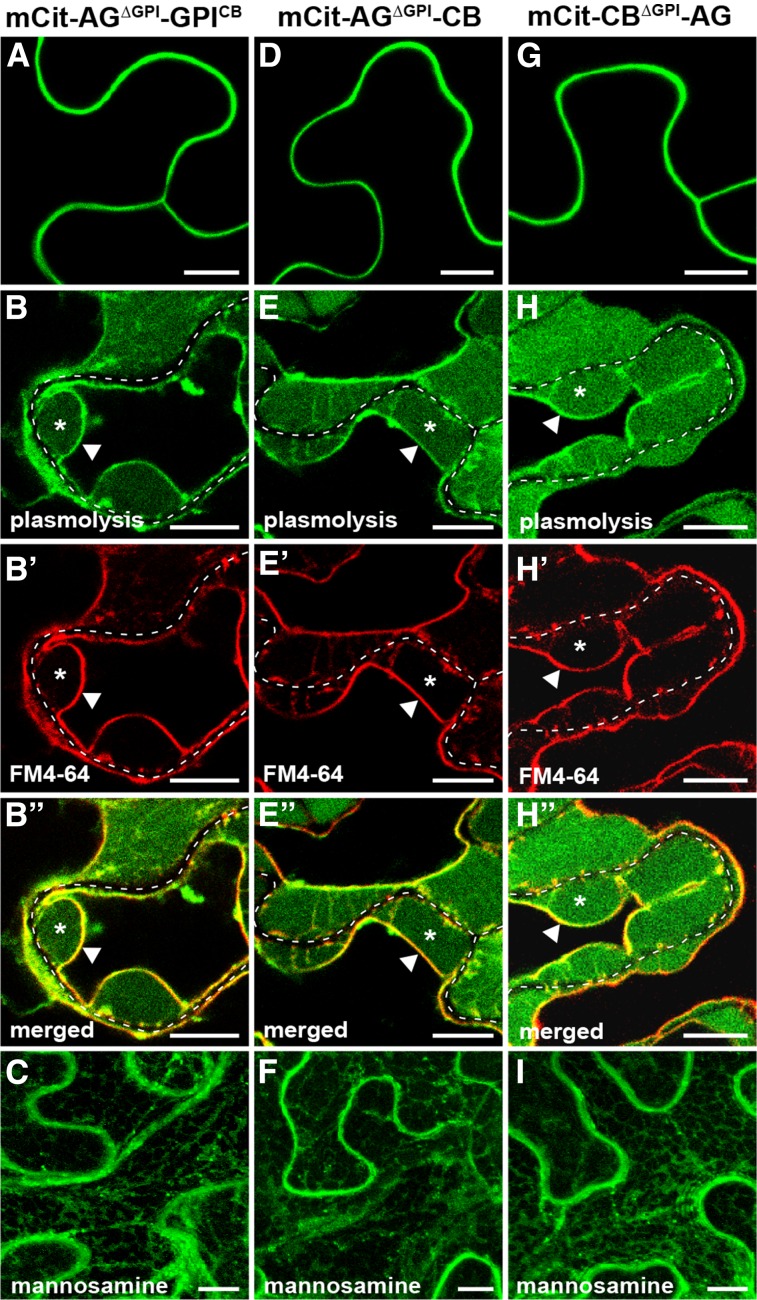
Figure 7.
Pd exclusion is dominant over Pd localization. Subcellular localization is shown for chimeric fusions between AGP4 and PDCB1 in transient expression in N. benthamiana. A to C, Localization of SP-mCitrine fused to AGP4 in which the GPI signal was replaced with that of PDCB1 (mCit-AGΔGPI-GPICB). D to F, Localization of SP-mCitrine fused to AGP4 in which the GPI signal was replaced with the full-length PDCB1 (mCit-AGΔGPI-CB). G to I, Localization of SP-mCitrine fused to PDCB1 in which the GPI signal was replaced with the full-length AGP4 (mCit-CBΔGPI-AG). A, D, and G show localization with no treatment; B to B′′, E to E′′, and H to H′′ show localization in plasmolyzed cells stained with FM4-64. In plasmolyzed cells, the dashed white line marks the cell wall, asterisks indicate the apoplastic space formed by the receding protoplast, and triangles indicate the position of the PM. C, F, and I show localization in the presence of the GPI biosynthesis inhibitor mannosamine. Bars = 10 µm.

