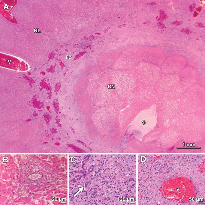
Figure 3:
Photomicrographs (H-E stain) of porcine liver tissue 7 days after treatment (with 415 mW). * = position of the laser fiber on each image. A, Photomicrograph (original magnification, ×10) shows that the treated area consisted of an area of CN surrounding the path of the laser fiber, surrounded by a region of fibrotic tissue (FZ) contraction. Residual areas of hemorrhage persisted in this concentric ring of fibrosis that appeared to encroach on the lesion from the periphery. A normal-appearing region (NL) was sharply delineated outside the treatment area, with an intact vein (v) in the field. B, High-power view (original magnification, ×400) from the CN region of the treated area shows ablation of hepatocytes and necrosis of the bile ducts and vessels within the portal triad. C, High-power view (original magnification, ×400) from the zone of fibrosis shows increased density of preexisting blood vessels that were viable after treatment, along with immature blood vessels (arrow) that are suggestive of neovascularization. The region is infiltrated with chronic inflammatory cells. D, Photomicrograph (original magnification, ×200) shows a portal triad that contains an intact blood vessel (v) in the periphery of the fibrotic zone is pictured, indicating sparing of the structure after treatment.

