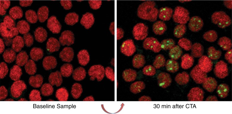
Figure 2b:
Schematic shows the study design. (a) Peripheral blood sample obtained before the patient underwent coronary CT angiography (CTA) provides baseline for comparison to repeat sample drawn 30 minutes after in vivo radiation exposure. A second tube of blood obtained prior to CT angiography is taped to the patient’s sternum for ex vivo radiation at the breast-tissue level with or without breast shielding (top right). (b) γ-H2AX foci induced by CT angiography, comparing baseline blood sample versus blood sample obtained 30 minutes after CT angiography. Green, γ-H2AX foci; red, DNA stained with propidium iodide.

