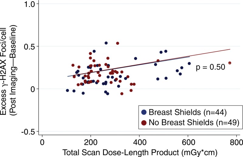
Figure 4:
Scatterplot shows blood samples of excess γ-H2AX foci that resulted from in vivo radiation during coronary CT angiography. Excess DNA double-strand-break levels measured by γ-H2AX immunofluorescence before and after coronary CT angiography demonstrated no difference in DNA damage between patients randomized to the use versus absence of breast shielding (P = .50 between groups).

