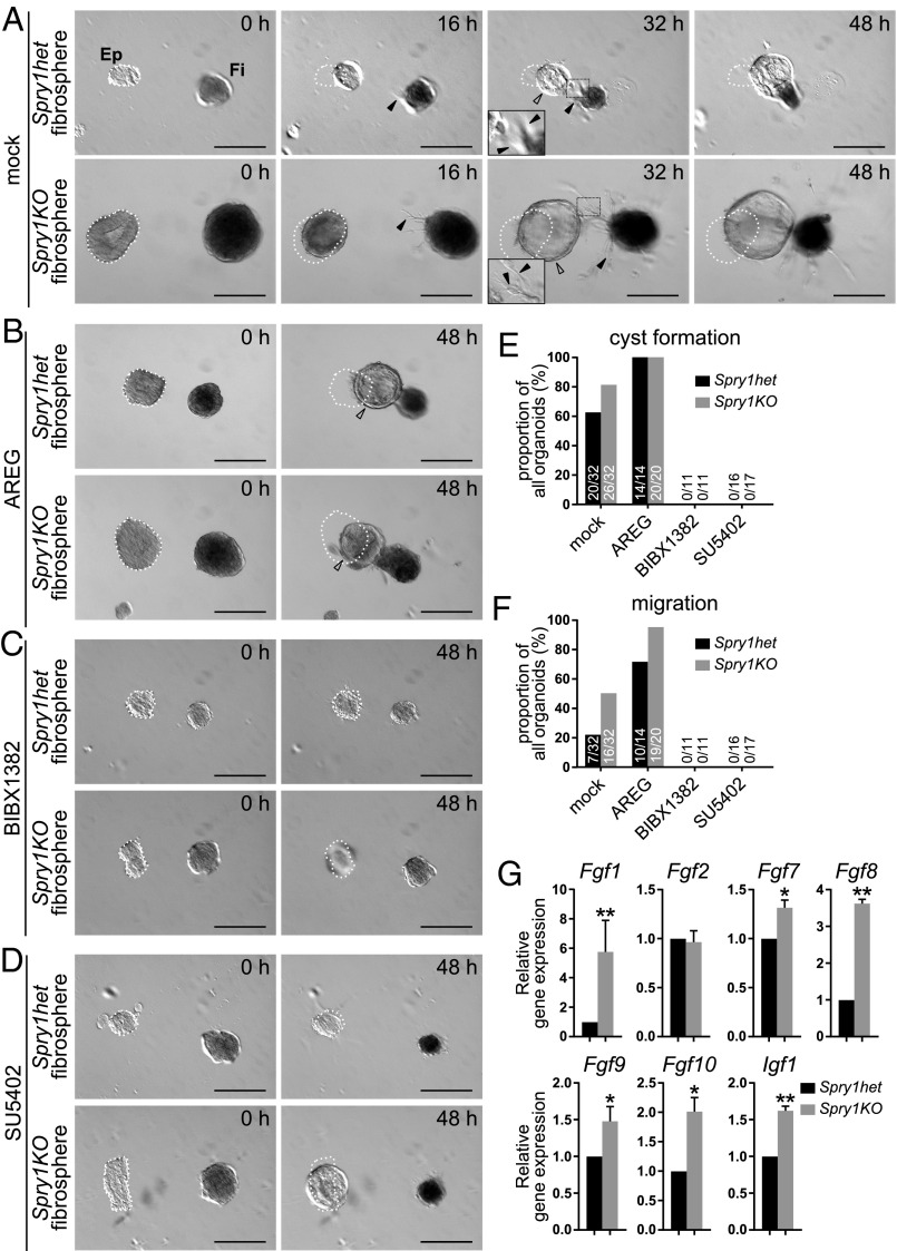
Fig. 3.
Mammary fibroblasts lacking Spry1 promote epithelial morphogenesis more strongly than normal in a paracrine fashion. (A–D) Time course of in vitro cocultures of wild-type epithelium (Ep; Left) with Spry1het or Spry1KO fibrospheres (Fi; Right) in the absence (A) or presence (B) of AREG stimulation, EGFR inhibitor BIBX1382 (C), or FGFR inhibitor SU5402 (D). (A) The epithelium formed a cyst (hollow arrowhead) and underwent directional collective migration toward fibrospheres, whereas fibrospheres formed spiky extensions made of fibroblasts (filled arrowheads), often in the direction of the epithelium. The images were recorded using time-lapse microscopy over 48 h, and the respective movies can be seen in Movies S1 and S2. (B–D) The ability of the epithelium to undergo cyst formation and collective migration was increased by AREG addition (B) and inhibited by EGFR inhibitor BIBX1382 (C) or FGFR inhibitor SU5402 (D). (Scale bars, 200 µm.) White dotted line indicates the original position of organoids at time 0 h. (E and F) Quantification of epithelial response to Spry1het and Spry1KO fibrospheres. Plots show proportions, as well as actual numbers of organoids, that underwent cyst formation (E) or migration (F) out of total number of organoids per condition. (G) Relative expression of candidate growth factor genes in serum-starved Spry1het and Spry1KO fibroblasts, normalized to Actb. Data shown are mean ± SEM (n = 3–4). Statistical analysis was performed using paired t test; *P < 0.05; **P < 0.01. Refer to Table S1 for primer sequences.

