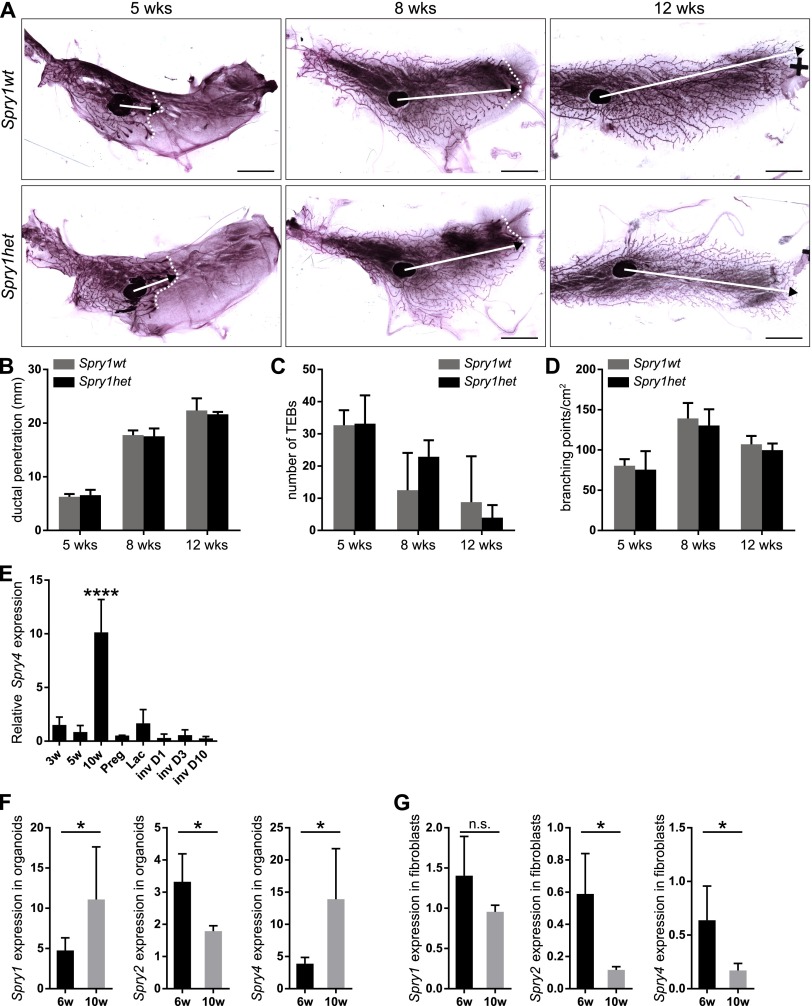
Fig. S1.
Whole-mount analysis reveals no difference between Spry1wt and Spry1het mammary glands. (A) Whole mounts of mammary glands from 5-, 8-, and 12-wk-old Spry1wt and Spry1het mice stained with carmine red. Images of parts of the mammary gland were merged in Photoshop (Adobe) to create a full view of the wholemount gland. Arrows indicate the extent of ductal penetration in the fat pad; dotted white line illustrates the epithelial invasion front. (Scale bars, 5 mm.) (B–D) Quantitative comparisons of ductal penetration (B), number of terminal end buds (TEBs) (C), and branching (D) between Spry1wt and Spry1het glands. Plots show mean ± SD (n = 3–5 litters/age). Statistical analysis was performed using two-way ANOVA and revealed no significant difference between Spry1wt and Spry1het mammary gland. (E–G) Expression of Sprouty genes in mammary gland. (E) Relative Spry4 expression during mammary gland development. RNA was isolated from mammary glands from virgin female mice at 3, 5, and 10 wk of age, and female mice on day 5 of pregnancy (Preg), day 1 of lactation (Lac), and days 1, 3, and 10 of involution (Inv). Graph shows mean ± SD (n = 3). Statistical analysis was performed using one-way ANOVA; ****P < 0.0001. (F and G) Relative expression of Sprouty genes in mammary epithelial organoids (F) and fibroblasts (G) from 6- and 10-wk-old mice. Graphs show mean ± SD (n = 3–7). Statistical analysis was performed using unpaired t test; *P < 0.05; n.s., not significant.

