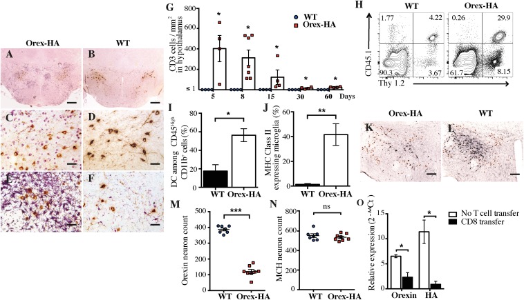
Fig. 2.
Orex-HA animals develop massive hypothalamic inflammation and marked orexin neuron loss after transfer of neo-self-antigen–specific CTLs. (A–D) Immunohistochemistry staining for CD3 (violet) and orexin-A (brown) in Orex-HA mice (A and C) and WT mice (B and D) 8 d after adoptive transfer of 3 × 10⁷ CTLs. (E and F) Immunohistochemistry staining of microglial (Iba1; violet) and orexin+ neurons (brown) in Orex-HA (E) and WT (F) mice. Representative results from four to seven mice per group are shown. [Scale bars: 200 μm (A and B) or 50 μm (C–F).] (G) Quantification of T cells in the hypothalamus of Orex-HA and WT mice at different time points after CTL injection. Results are expressed as mean ± SEM of four to seven mice per group for each time point. (H) Representative FACS plots of brain-infiltrating cells from Orex-HA and WT littermates on day 8 after CTL transfer. (I and J) The proportion of CD11c+ among CD45high CD11b+ cells (I) and the expression of MHC class II molecules on CD45dim CD11b+ Thy1.2− cells microglia (J) were assessed by flow cytometry. Results are expressed as mean ± SEM of six to eight mice per group from three independent experiments. Statistical analyses were performed by using the Mann–Whitney u test. *P < 0.05; **P < 0.01, comparing the Orex-HA group with the respective WT controls. (K and L) At 60 d after CTL transfer, immunohistochemistry staining for orexin+ neurons (black) and MCH+ neurons (brown) in Orex-HA animals (K) and WT mice (L). [Scale bars: 125 μm (K and L).] (M and N) Quantification of orexin+ (M) and MCH+ (N) neuronal cell bodies in the hypothalamus of Orex-HA mice compared with WT animals 60 d after CTL transfer. Results are expressed as mean ± SEM of seven or eight mice per group from two independent experiments. (O) Expression of orexin and HA mRNA in the hypothalamus of Orex-HA mice that received neo-self-antigen–specific CTLs or have been left untreated. Results are expressed as mean ± SEM of four or five mice per group from two independent experiments. Statistical analyses were performed by using the Mann–Whitney u test. *P < 0.05; **P < 0.01; ***P < 0.001. ns, not significant.

