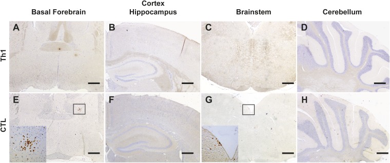
Fig. S3.
Mild or no T-cell infiltration is detected outside the hypothalamus in Orex-HA mice injected with HA-specific Th1 or CTL. Immunohistological staining of brain sections of Orex-HA mice at day 8 after Th1 (A–D) or CTL transfer (E–H) to detect T-cell infiltration (CD3, brown) in extrahypothalamic regions including basal forebrain (A and E), cortex, and hippocampus (B and F), brainstem (C and G), and cerebellum (D and H) is shown. Insets highlight rare CD3+ cells in the brain parenchyma. Nuclear counterstaining was performed with hematoxylin (blue). Representative results from four or five mice per group are shown. (Scale bars: 200 μm.)

