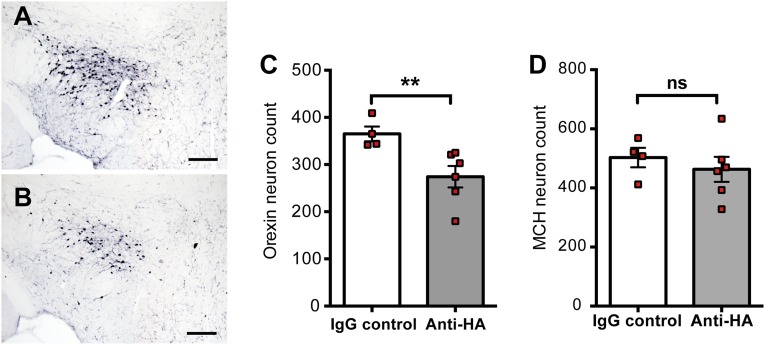
Fig. S6.
Anti-HA antibodies induce mild loss of orexin+ neurons in the context of Th1 inflammatory lesions. Enumeration of orexin+ neurons 60 d after transfer of 3 × 10⁷ Th1 cells in Orex-HA mice by Immunohistochemistry. (A and B) Mice received either 200 μg of control IgG (A) or anti-HA antibodies (B) every 2 d, from day 5 to day 15 after Th1 transfer. (Scale bars: 150 μm.) (C and D) Quantification of orexin+ (C) and MCH+ (D) neuronal cell bodies in the hypothalamus of Orex-HA mice that received the control IgG (white bars) or the anti-HA antibodies (gray bars). Results are expressed as mean ± SEM of four to six mice per group. Statistical analysis was performed by using the Mann–Whitney u test. **P < 0.01. ns, not significant.

