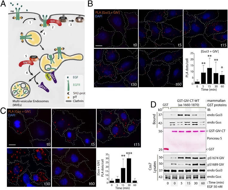
Fig. 1.
Upon EGF stimulation GIV binds Gαi3 and Gαs in a sequential manner. (A) Schematic summarizing GIV’s interactions with Gαi3 and Gαs at various steps in EGFR signaling and trafficking (modified from ref. 9). Upon EGF stimulation, GIV is recruited to the PM (step 1), assembles an EGFR–GIV–Gαi3 complex at the PM (step 2), activates Gαi3 (step 3), and prolongs the association of EGFR with the PM. Upon internalization, EGFR traffics to APPL endosomes (step 4) and then to EEA1 endosomes where GIV binds inactive Gαs (step 5), promoting dissociation of EEA1 and endosome maturation to multivesicular endosomes (step 6) to facilitate EGFR down-regulation, thus shutting off endosome-based proliferative signaling. (B and C) Serum-starved (0.2% FBS, overnight) HeLa cells were stimulated with EGF at the indicated time points and were fixed and analyzed for interactions between GIV and Gαi3 (B) or GIV and Gαs (C) by in situ PLA (red). Nuclei are stained with DAPI (blue). Cell boundaries were traced with interrupted lines by superimposing bright-field microscopy images. Results are expressed as mean ± SEM; n = 3. (Scale bar, 15 µm.) (Lower Right) The number of PLA dots per cell was quantified from a total of 25–30 cells per experiment. (D) Cos7 cells expressing GST-GIV-CT (amino acids 1660–1870) were stimulated with EGF at the indicated time points and were lysed and incubated with glutathione-Sepharose beads. (Top) Bound proteins were analyzed for endogenous Gαi3 and Gαs by immunoblotting (IB). (Middle) Equal loading of GST and GST-GIV-CT proteins was confirmed by Ponceau-S staining. (Bottom) Cell lysates were analyzed for pS1674-GIV, pS1689-GIV, Gαi3, and Gαs by immunoblotting and were quantified using LI-COR Odyssey (Fig. S1). *P < 0.05, **P < 0.01, ***P < 0.001.

