Fig. 4.
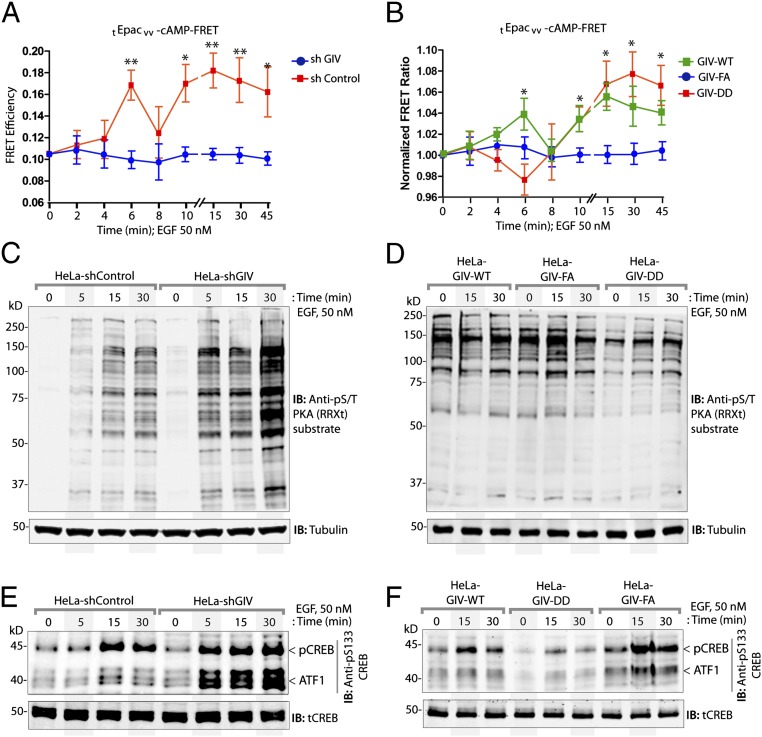
GIV inhibits the Gαs–GTP→cAMP→PKA→CREB pathway. (A and B) Control (shControl) and GIV-depleted (shGIV) HeLa cells (A) or GIV-WT, GIV-DD, or GIV-FA Hela cell lines (B) expressing the tEpacvv-cAMP FRET probe were serum starved (0.2% FBS) and subsequently stimulated with 50 nM EGF and analyzed for ratiometric FRET imaging using a confocal microscope (increase in cAMP = loss of FRET, and vice versa). Graphs display the change in FRET efficiency (y axis) over time (x axis). Results are expressed as ± SEM; n = 3. *P < 0.05, **P < 0.01. (C and D) Serum-starved control (shControl) or GIV-depleted (shGIV) HeLa cell lines (C) or GIV-depleted HeLa cells stably expressing GIV-WT, GIV-DD, or GIV-FA (D) were stimulated with 50 nM EGF at the indicated time points before lysis. Equal aliquots of whole-cell lysates were analyzed for PKA activity by immunoblotting using anti–phospho-serine/threonine–PKA substrate-specific antibody and tubulin. (E and F) HeLa cell lysates in C and D were analyzed for pCREB, phospho-ATF1, and total CREB (tCREB) by immunoblotting and were quantified by band densitometry (Fig. S5 A and B).

