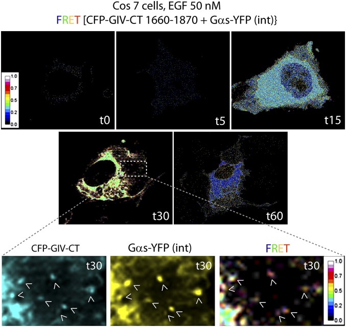
Fig. S2.
Spatiotemporal dynamics of GIV–Gαs interaction after EGF stimulation, as determined by FRET imaging. Cos7 cells were first cotransfected with internally tagged Gαs-YFP [Gαs-YFP(int)] and a previously validated functional CFP-tagged GIV-CT (amino acids 1660–1870) probe which retains key properties of full-length GIV (i.e., the ability to couple G-protein signaling to ligand-activated growth factor receptors). Cells were starved overnight (0% FBS), stimulated with EGF, and analyzed for FRET by confocal live-cell imaging at different time points (t0–t60). Representative images from different time points are displayed. Images show the intensities of acceptor emission triggered by energy transfer in each pixel. Maximum interaction between GIV and Gαs, as determined by increased FRET, occurred at t30 in a perinuclear compartment and on endosome-like structures (Insets). (Magnification, 63×.)

