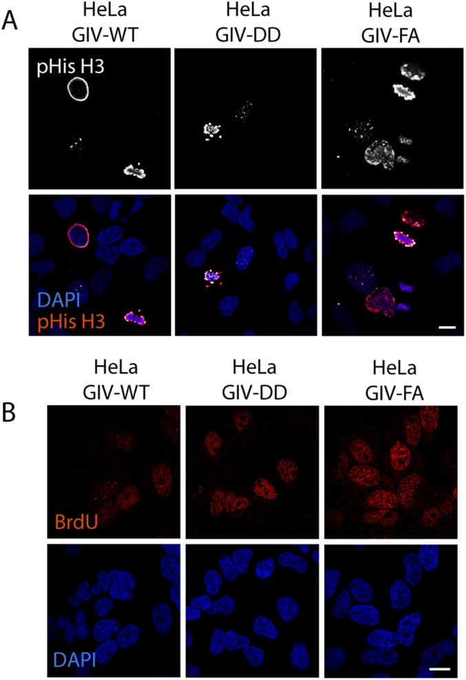
Fig. S7.
GDI-proficient HeLa GIV-DD cells proliferate more slowly than the GDI-deficient HeLa GIV-FA cells. (A) GIV-depleted HeLa cells expressing GIV-WT or DD or FA mutants were grown in 2% (vol/vol) FBS before fixation. Fixed cells were stained for phosphorylated (S28)-histone H3 (red) and DAPI (to stain the nucleus; blue) and were analyzed by confocal microscopy. Images of representative fields are shown. (Scale bar, 10 µm.) Quantification of positively stained cells is displayed as bar graphs in Fig. 5D. (B) HeLa cell lines in A were grown in 2% (vol/vol) FBS overnight, incubated with BrdU for 30 min, fixed and stained for anti-BrdU mAb (red) and DAPI (to stain the nucleus; blue), and analyzed by confocal microscopy. Images of representative fields are shown. (Scale bar, 10 µm.) Quantification of positively stained cells is displayed as bar graphs in Fig. 5E.

