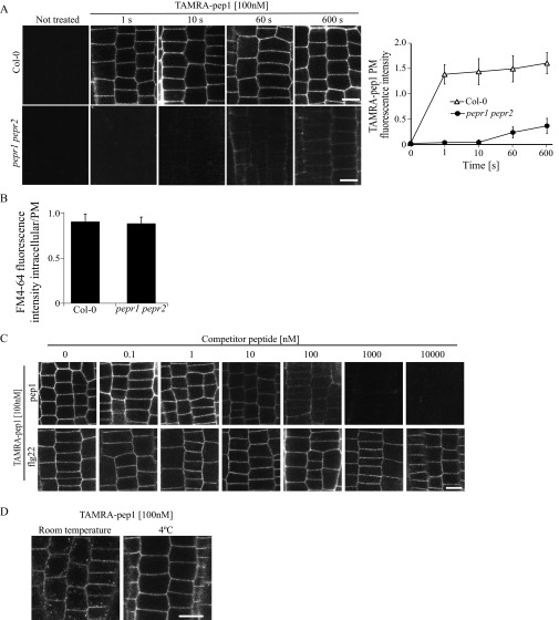
Fig. S3.
PM labeling of TAMRA-pep1. (A) PM of Arabidopsis WT (Col-0) and pepr1perp2 double-mutant root meristem epidermal cells labeled within seconds after application by TAMRA-pep1. The 5-d-old seedlings were treated with TAMRA-pep1 (100 nM) for different time points, washed three times, and then imaged. (Right) Quantification of fluorescence intensity in the PM (n = 48). Error bars indicate SD. (B) Quantification of the FM4-64 signal intensity in Col-0 and pepr1perp2 double mutant. Values represent the intracellular/PM signal intensity ratio in root meristem epidermal cells of 5-d-old seedlings treated with FM4-64 (4 µM, 5 min, three washouts) and kept in the presence of 100 nM pep1 for another 90 min (n >32). Error bars indicate SEM. No statistically significant difference (*P ≤ 0.01, Student’s t test) was found. (C) pep1, but not flg22, is able to compete with TAMRA-pep1. Five-day-old Arabidopsis seedlings were incubated with various concentrations of pep1 or flg22 for 5 min, then treated with TAMRA-pep1 for 10 s, washed, and imaged. The fluorescence intensity of the PM is quantified in Fig. 1D. (D) Temperature-dependent internalization of TAMRA-pep1. Five-day-old Arabidopsis seedlings were incubated at 4 °C for 2 h before treatment with TAMRA-pep1 (100 nM, 10 s), washed, and imaged after incubation for 40 min at 4 °C. The same TAMRA-pep1 treatment was done with seedlings at room temperature. n, number of cells analyzed. (Scale bars: 10 µM.)

