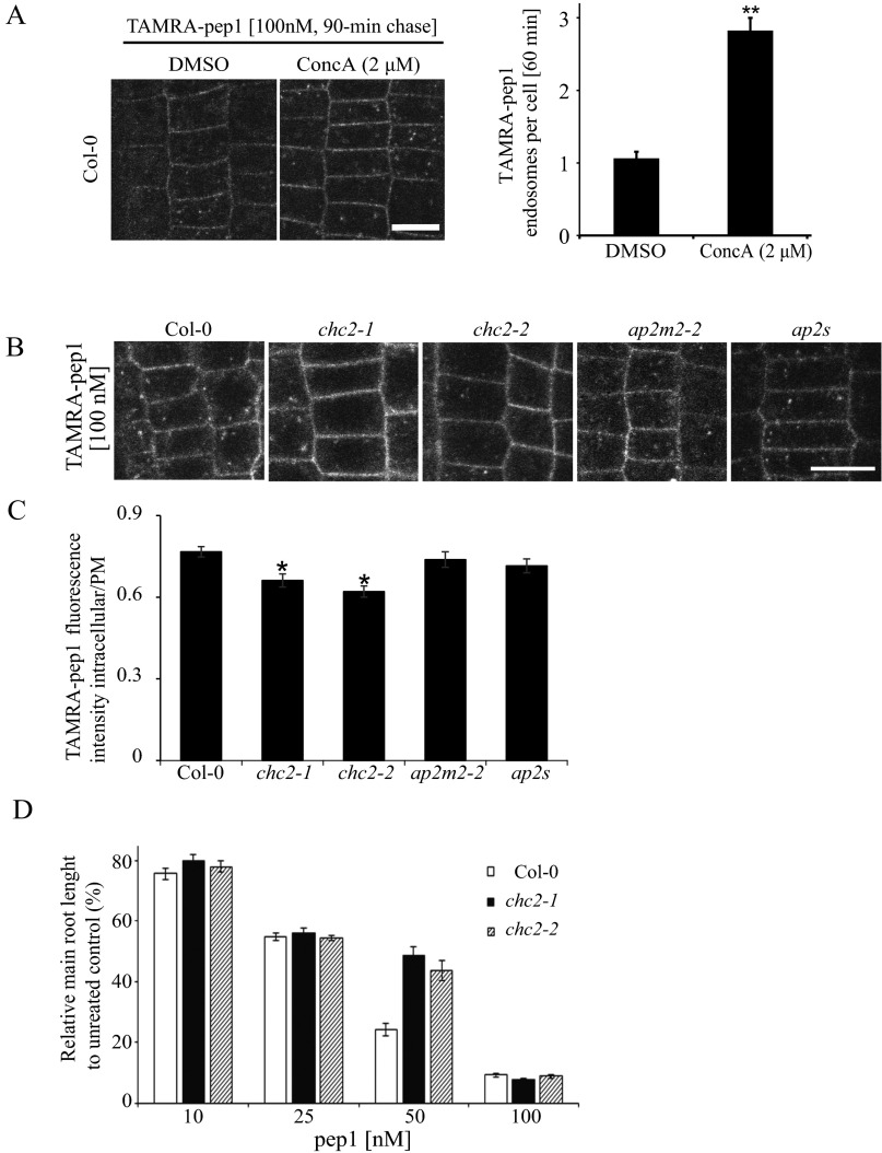
Fig. S8.
Clathrin-dependent TAMRA-pep1 internalization and AtPep1-mediated responses. (A) Impaired activity of vacuolar H+-ATPase by the presence of ConcA slightly delaying the pass of TAMRA-pep1 to the vacuole. Quantification of the number of TAMRA-pep1 endosomes per cell (n = 36). n, number of cells analyzed. Error bars indicate SEM. **P ≤ 0.001 (Student’s t test) relative to Col-0. The 5-d-old seedlings were pretreated with ConcA (2 µM, 30 min), then treated with TAMRA-pep1 (100 nM, 10 s), washed three times, and kept in the presence of ConcA (2 μM) until imaging (60-min chase). DMSO served as a control. (Scale bars: 10 μm.) (B) TAMRA-pep1 uptake in WT Arabidopsis (Col-0; control) and chc2-1, chc2-2, ap2m-2, and ap2s homozygous mutants. The 5-d-old seedlings were treated with TAMRA-pep1 (100 nM, 10 s), washed three times, and imaged after a 40-min chase. (C) Quantification of the TAMRA-pep1 uptake in B. Graphs illustrate the intracellular/PM fluorescence intensity ratio (n >45). n, number of analyzed cells. Error bars indicate SEM. *P ≤ 0.01 (Student’s t test) relative to Col-0. (Scale bar: 10 µm.) (D) Root growth analysis of Col-0, chc2-1, and chc2-2 seedlings grown in the presence of various concentrations of pep1. Data are the sum of two biological repeats, and the main root length is presented as relative to the mock control. Error bars indicate SEM.

