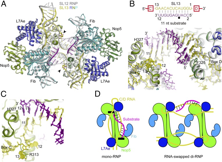
Fig. 3.
Structure of SL13 RNP bound to 11-nt substrates. (A) Structural alignment of SL13 RNP bound with 11-nt substrates and SL12 RNP bound with 10-nt substrates. The SL12 RNP structure is colored in silver. The SL13 RNP structure is colored by molecule; the two Nop5 subunits are in dark green and light green, L7Ae is in blue, fibrillarin (Fib) is in cyan, C/D RNA is in yellow, and substrate RNA is in purple. The looped-out guide nucleotide 13 is marked with an arrowhead. (B) Conformation of guide-substrate duplexes in the aligned SL12 RNP and SL13 RNP structures. Fibrillarin is omitted for clarity. Proteins are shown as ribbons; RNAs, as sticks. Base pairing interactions between the guide and the substrate are displayed at the top. The target site is marked by a black circle. Nucleotides in spacers and substrates are numbered by their distances to box D. (C) Close-up view of the SL13 RNP structure at the box C' side. (D) Cartoons of C/D mono-RNP and RNA-swapped di-RNP. Black boxes indicate the structural elements that cap the guide-substrate duplex in the mono-RNP model.

