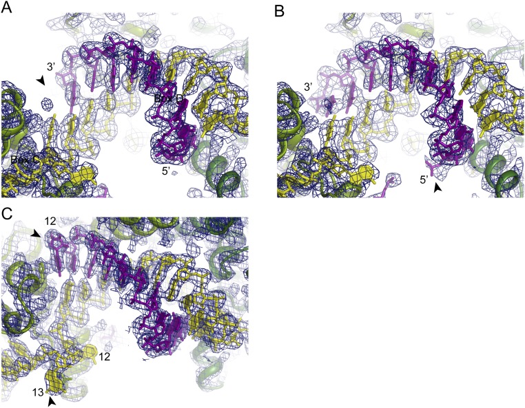
Fig. S1.
Electron density map of C/D RNP structures. The 2Fo–Fc electron density map contoured at the 1.5σ level is displayed for the SL12 RNP structure bound to the 9-nt (A) or 13-nt (B) substrates and the SL13 RNP structure bound to the 11-nt substrate (C). The determined structures are overlaid on the map. Substrate RNAs are shown in purple, C/D RNAs are in yellow, and two Nop5 subunits are in light green and dark green. The SL12 RNP structures in A and B have the same orientation. Important features in each map are marked with arrowheads. In A, the arrowhead indicates a missing density at position 11 for the 9-nt substrate. In B, the arrowhead indicates extra density for a phosphate group from the 5′ extension of the 13-nt substrate. In C, the arrowheads indicate the unpaired substrate nucleotide at position 12 and the unpaired spacer nucleotide at position 13.

