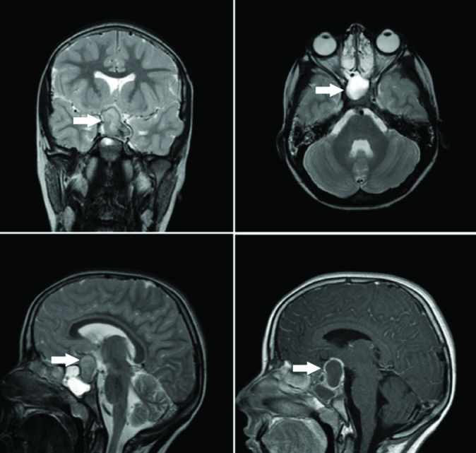
Figure 2.

Mass lesion with a size of approximately 3×3,5×3 cm which filled the base of the sella, compressed the left optic nerve and the optic chiasma on the left with regular borders and contrast enhancement following intravenous injection of contrast material
