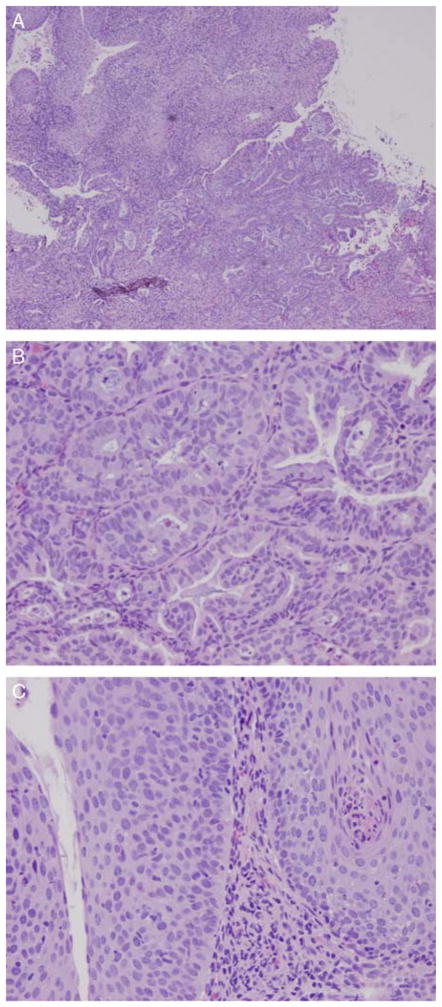
FIG. 1.
(A) A low-power magnification of the vaginal biopsy hematoxylin and eosin section revealed a well-differentiated adenocarcinoma, and the overlying squamous mucosa showed high-grade vaginal intraepithelial neoplasia (10×). (B) The adenocarcinoma component consisted of back-to-back glands with slight nuclear atypia and few mitotic figures (40×). (C) The squamous mucosa overlying the adenocarcinoma shows a high-grade vaginal dysplasia in which the cells lacked normal maturation and reached the entire thickness of the squamous epithelium. Koilocytosis resulting from human papilloma virus effect are also present.

