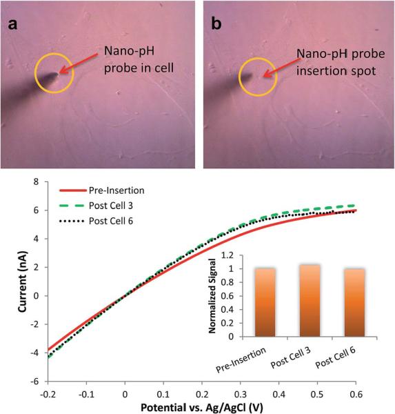
Fig. 5.
Representative micrographs of (A) a nano-pH probe inserted to a MDA-MB-231 and (B) after retraction of the probe. Cells did not show any morphological changes and stayed intact over the course of insertion and measurement, and survived after retraction. (C) In vitro reusability of the nano-pH probes. Linear sweep voltammograms of regenerating baseline of the same nano-pH probe after the third and sixth cell interrogation in 0.1 M PBS (pH 7.0).

