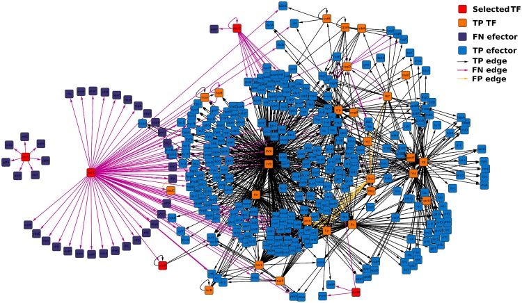
Fig 5. Merged sub-network of the TFs with the lowest RGD using suspension network at 15 hours as reference.
The TF encoding genes identified using their RGD are colored in red; other TFs are colored in orange and they are named TP because their expression was detected in the two compared networks; effector genes, those that do not code TFs are colored in purple if their expression was detected only in the reference network (FN nodes) and in blue if their expression was detected in both networks; with respect to the edges, they are colored in black if they were present in both networks (TP edges), light purple if they were found only in the reference network (FN edges) and yellow when they were detected only in the compared network (FP edges). The same image but with TP edges removed is shown in S5 Fig.

