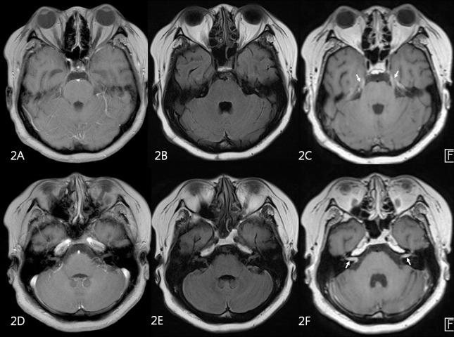
Fig 2. MR images of 58 year-old male patient with advanced gastric cancer.
2D contrast-enhanced T1-weighted GRE (A,D) and 2D FLAIR (B,E) were negatively interpreted by both faculty and trainee raters. On 3D contrast-enhanced T1-SPACE (C,F), leptomeningeal enhancement along trigeminal nerves (arrows on C) and internal auditory canals (arrows on F) was seen.

