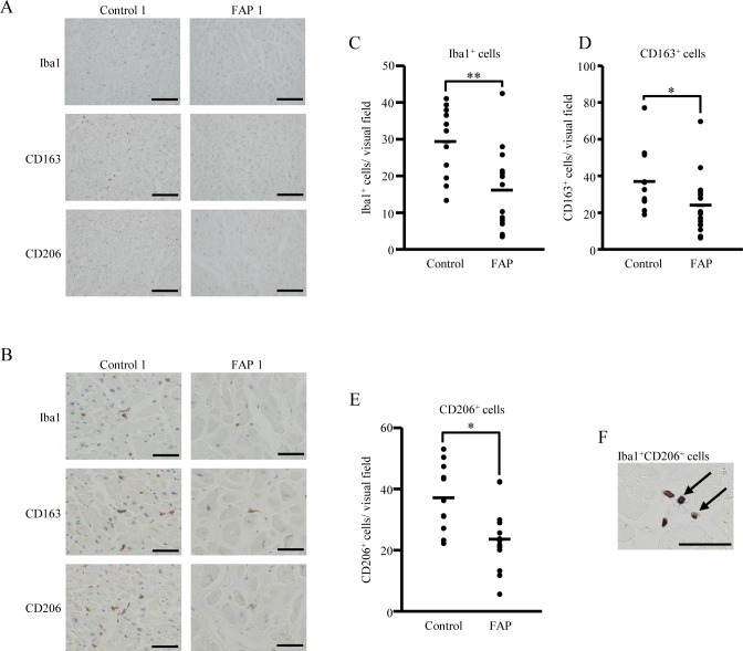
Fig 2. Lower number of tissue-resident macrophages in FAP ATTR V30M patients.
(A, B) Heart tissue (FAP ATTR V30M patients, n = 16; control patients, n = 11) was stained with macrophage-related (Iba1) and inhibitory macrophage (CD163 and CD206) markers by immunohistochemistry. All slides shown are representative of each group. Lower (A) and higher (B) magnification views are shown. (C-E) Five visual fields in each stained section were randomly chosen, and the number of Iba1, CD163, and CD206-positive cells counted by two independent observers. Graphs show the average count number per visual field for each marker: Iba1 (C), CD163, (D) and CD206 (E). Repeated count immunostained cells were analyzed using the generalized Poisson mixed model, with *p < 0.05 and **p < 0.01 indicating significant differences. (F) Double immunohistochemical staining of Iba1 and CD206 in heart tissue of FAP ATTR V30M patients. Black arrows show double-immunostained cells. Bars indicate 200 μm (A) and 50 μm in (B, F).

