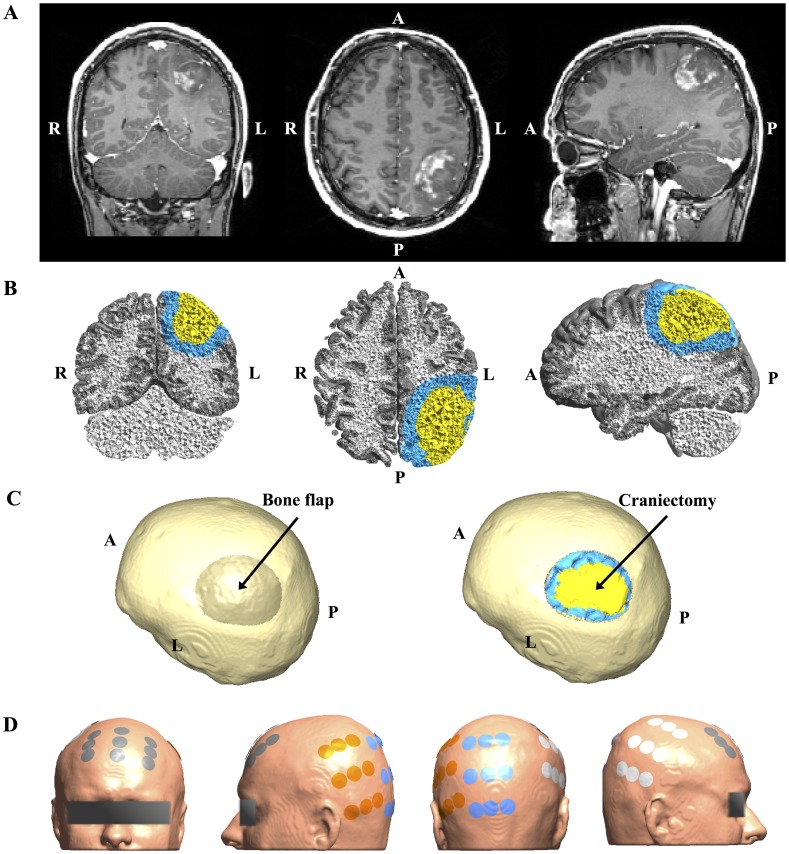
Fig 1. MRI data from study Subject 1 and corresponding 3D head model.
A. Coronal (left), axial (middle) and sagittal (right) views of original Gadolinium enhanced T1 MRI patient data showing left parietal glioblastoma (radiological orientation). B. Volume reconstruction of gray matter (gray), white matter (white), tumor tissue (yellow), and a peritumoral region (blue). C. Surface reconstruction of patient skull rotated to present the craniectomy boneflap outlined as a darkened area above the tumor (left). The rightmost image shows the same view, but with the bone flap removed to display the underlying tumor and peritumoral region. D. Surface reconstruction of the head model showing the optimized electrode layout used for simulation (NovoTAL ™). Electrodes are paired orange with white and gray with blue.

