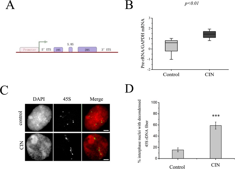
Fig 2. The rRNA level was increased in human cervical cancer samples.
(A) Schematic representation of a human 45S rRNA gene. (B) The box plot reveals the relative levels of the rRNA in CIN as compared with the non-tumor tissue. Significance between normal (control) and tumor (CIN) samples was determined using the sign test (the P-value is less than 0.001). (C) Cancer caused aberrant 45S rDNA signal patterns in nuclei. FISH of nuclei with 45S rDNA probes showed spot signals in non-cancer tissues and fiber-like threads unraveled from compacted states in CIN tissues. Bar = 10 μm. (D) Percentages of interphase nuclei with decondensed 45S rDNA fibers in CIN or non-tumor tissues, respectively. Number of evaluated nuclei in each group was 250. Data are expressed as *P<0.05, ***P<0.01, measured by the t-test.

