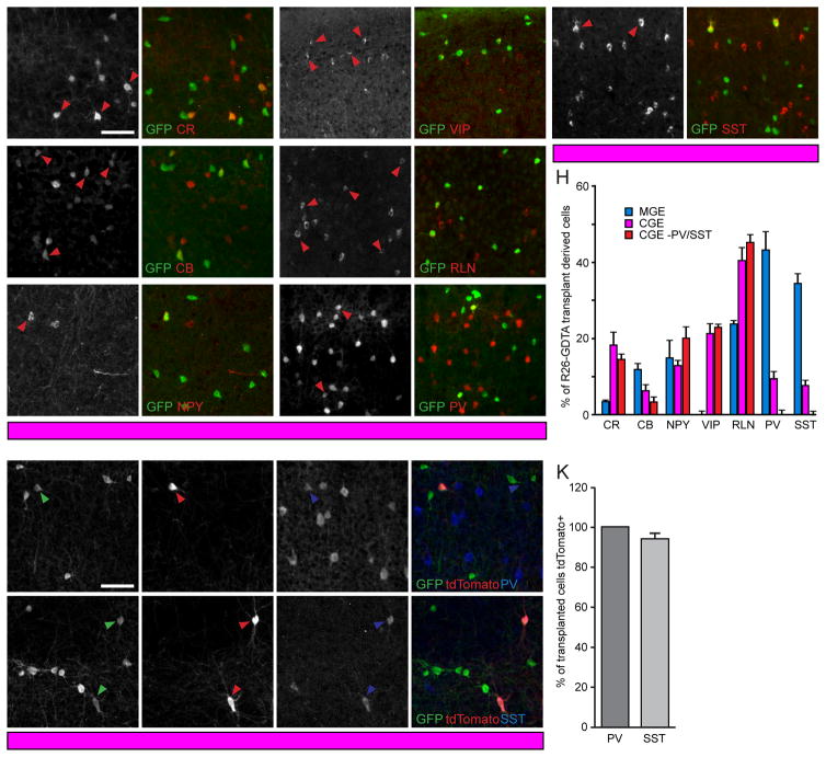
Figure 2. PV and SST Neurons In CGE Transplants Are MGE-Derived.
(A–G) Visual cortex coronal sections of CGE transplant recipients at 35DAT stained for GFP (A′–G′), CR (A,A′), CB (B,B′), NPY (C,C′), VIP (D,D′), RLN (E,E′), PV (F,F′) and SST (G,G′). Arrowheads identify double-labeled cells. Scale bar: 100 μm.
(H) Percent of transplanted GFP+ neurons immunoreactive for subtype markers in A–G found in R26-GDTA MGE (n=3 mice), R26-GDTA CGE (n=5 mice), and PV-Cre;SST-Cre;R26-GDTA CGE (n=3 mice) recipients at 35DAT. Error bars represent SEM. Significance calculated using Bonferroni corrected t-tests.
(I–J) GFP, tdTomato, PV, and SST staining at 35DAT illustrating the presence of Nkx2.1 lineage PV and SST interneurons in CGE transplants. Arrowheads identify triple-labeled cells. Scale bar: 100 μm.
(K) Percent of transplanted PV and SST interneurons which express tdTomato in anatomically isolated CGE transplants. Error bars represent SEM (n=3 mice).

